Heads Will Turn: How Neck Muscles Supported A Major Evolutionary Transition (Fixations #3)
In our latest Fixations post, we explore how evolution built upon existing muscle groups to craft a mobile neck during the water-to-land transition.
Introduction
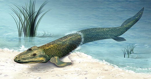
When you think of evolution, you might picture that iconic image of hominids emerging from lesser apes, but do you ever think about what came before there were land-dwelling animals? What about how animals got onto land in the first place? Lucky for us, scientists have been working on this question for decades by characterizing a 400 million year old evolutionary event known as the water-to-land transition. This event marked the emergence of the tetrapods–a group of four-legged vertebrates that would eventually give rise to humans. To enable such a dramatic change in lifestyle and environment, several morphological changes occurred in this group that allowed them to adapt to land. One of these you may be familiar with through the fossil Tiktaalik: the fin-to-limb transition. Discovered by Dr. Neil Shubin, Dr. Edward Daeschler, and their colleagues in 2006, the fossil Tiktaalik rosaeae provided the first morphological evidence of this transition. More specifically, the tetrapod possessed fins with shoulder, elbow, and even partial wrist components.
Another morphological change that enabled the water-to-land transition that is often less thought of, but just as important, is the transition of an immobile head to one that can freely rotate and assess the environment. Much like developing a limb from a fin, the ability to go from not moving your head at all to moving it independent of your body requires a dramatic anatomical change. Additionally, it’s not about just physically moving your head, but also supporting the functional structures that reside in your neck, like your pharynx (a muscle that helps with feeding and breathing), larynx (a hollow tube that acts as your voice box), and the hyoid bone (which supports these other structures). As a result, vertebrates today possess four complex networks of muscle in their necks: 1) the pharyngeal and laryngeal muscles, 2) hypobranchial muscles (which connect to the hyoid bone), 3) epaxial and hypaxial muscles (which surround the vertebrae), and 4) cucullaris-derived muscles (which overlay other neck musculature). The first two contribute to processes like food intake, respiration, and vocalization, while the latter two are involved with mobility and locomotion.

In order to understand how these muscles came to support this evolutionary transition, however, it’s important to understand how they develop and how this development varies across different vertebrates. This is just what researchers from the Institut Pasteur’s Tajbakhsh lab, the National Museum of Natural History France, and the University of Colorado School of Medicine set out to do in their recent study, which both characterizes the developmental origins of the cucullaris-derived muscles in the zebrafish and uses multiple vertebrate models to understand how different muscle groups have changed over the course of evolution. Using a mix of lineage tracing techniques (figuring out what cells gave rise to a cell population of interest during development), molecular techniques, and high-powered imaging techniques, this study sheds light not only on how early neck muscles emerged in evolutionary history, but also how different muscle groups have changed throughout the course of it.
Main Findings
To figure out the identity of a muscle, you typically need to know three things about it: what embryonic tissue it comes from, what nerves connect to it, and what bones the muscle itself connects to. Accordingly, the authors of this study decided to tackle each of these questions to determine how different parts of the mesoderm–or, the embryonic layer that gives rise to all muscle in an organism during development–contributed to the eventual emergence of a mobile neck in the tetrapods.
To start, the authors wanted to understand whether the muscles that connect the head and trunk were homologous between zebrafish and tetrapods. More specifically, do these muscles have similar embryonic origins and are they innervated by the same nerves? To answer this question, they decided to use transgenic zebrafish reporters–in other words, zebrafish that have had a fluorescent protein attached to a gene of interest to visualize that gene’s activity–targeting two different mesoderm populations: the somitic mesoderm and the cardiopharyngeal mesoderm.
Beginning with the somitic mesoderm reporter, which uses the gene pax3a, the authors analyzed both when and where they detected expression of the reporter during the first five days of zebrafish development. In doing so, they aimed to track development of the head-and-trunk muscles (HTM) and the emergence of what will eventually become the cucullaris muscles. Their analysis showed that, while pax3a expression was associated with the hypaxial muscles throughout development, it was not associated with the cucullaris or other muscles. The authors next turned to the cardiopharyngeal mesoderm, using a genetic lineage tracing tool driven by the gene tbx1 to map the muscles emerging from this mesoderm population during the first five days of zebrafish development. They found that the cucullaris muscle fibers showed successful lineage labeling, while other muscle groups–such as the hypaxial muscles–did not show any labeling. Together with their pax3a analysis, the authors concluded that the cucullaris muscles emerge from the cardiopharyngeal mesoderm instead of the somitic mesoderm in the zebrafish–a finding that has also been confirmed in previous mouse studies.
Having answered the question of the muscles’ embryonic origins, the authors next wanted to investigate the innervation pattern of these muscles. For these experiments, they continued to use the zebrafish in addition to using the axolotl as a tetrapod comparison. Using a technique called antibody staining to visualize the nerves and muscles as well as a form of microscopy called confocal microscopy, they created a 3D rendering of a zebrafish larva to better understand how the different nerves connect to their muscles of interest (Figure 3, below). Through this method, they observed that the developing cucullaris muscle was located close to the vagus nerve and eventually became connected to it at a later stage of development. These results were also recapitulated in the axolotl larvae, which, when taken together with the genetic lineage tracing and anatomical results, seem to indicate the presence of a cucullaris muscle homologue in zebrafish. In other words, prior to the water-to-land transition, bony vertebrate fish already had the developmental ground plans for the muscles that would eventually facilitate this transition.

Naturally, this raises the question of how the somitic and cardiopharyngeal mesoderm have changed throughout the course of vertebrate evolution, both before and after the water-to-land transition. To understand the HTM rearrangements that have characterized jawed vertebrate evolution, the authors decided to use a technique called micro-CT, which uses X-rays to gather 3D morphological data on internal structures. They drew upon a wide variety of vertebrates for their study, using the following pairs to elucidate differences in the hypobranchial, laryngeal, and cucullaris-derived muscles: the zebrafish and the bichir Polypterus senegalus (ray-finned fishes, the latter of which has a separation between its skull and shoulder bone), the African coelacanth and the African lungfish (lobe-finned fishes, which also have a skull and shoulder bone separation), the axolotl Ambystoma mexicanum and the emperor newt (salamanders that have short necks with limited mobility, one aquatic and one terrestrial, respectively), and the green anole lizard (a terrestrially adapted vertebrate with a flexible long neck), as seen in Figure 3 below.
Through their analyses, the authors determined that both groups of fishes–the ray-finned and the lobe-finned–had more limited hypobranchial and cucullaris muscles compared to the tetrapods. More specifically, certain parts of these muscles were variably missing among the fish; for example, the coelacanth did not have cucullaris-derived muscles, while the zebrafish and lungfish did not have a specific component of the hypobranchial muscle. Interestingly, though, the bichir, coelacanth, and lungfish all showed evidence of laryngeal musculature. Not only that, the laryngeal muscles in the bichir and lungfish actually bore resemblance to those of the axolotl, one of the salamanders included in the study. Similar to what we saw with the cucullaris muscles, this observation suggests that the laryngeal muscle structure of these fish is closer to that of terrestrial vertebrates despite their lack of a mobile neck.
Turning to our salamanders, the authors noted that both the axolotl and the newt exhibited differences in multiple types of musculature. For example, the axolotl’s laryngeal muscles had become associated with laryngeal cartilages located close to the esophagus. More importantly, however, they observed that the newt had a more extensively developed laryngeal musculature and that its cucullaris-derived musculature and hypobranchial musculature had extended onto other structures, suggesting that the terrestrial transition molded these muscles into the configuration needed to support an, albeit somewhat limited, neck. Lastly, in the green anole lizard, the authors saw the continuation of this trend in two ways: first, through additional extensions (or, making more connections to other structures) of the cucullaris-derived musculature, and second, through the association of the laryngeal musculoskeletal system to the hypobranchial musculature. Using these findings, the authors constructed a phylogenetic tree where they illustrate the different losses and expansions of these groups over time, which can be seen below.

Taken together, this study first sets out to better characterize the embryonic origins of the cucullaris-derived muscles in the zebrafish, before eventually expanding to assess how different muscles–namely, the laryngeal, hypobranchial, and cucullaris-derived muscles–have changed throughout the evolution of the tetrapods. In addition to confirming the cardiopharyngeal mesoderm as the tissue that eventually gives way to the cucullaris-derived muscles, the authors also provide evidence of a cucullaris muscle homologue in the zebrafish, showing that the foundations for the muscles needed to support a neck were present before the water-to-land transition. Then, by using micro-CT methods on multiple vertebrate samples, the authors show how the three muscle groups have changed, expanded, or added connections over the course of evolutionary time, giving us a better perspective of how these muscles came to support the necks we take for granted today.
The Takeaways
In uncovering how these different muscle groups have changed over the course of evolution, particularly as it pertains to the water-to-land transition, this work serves as a case study for understanding a much broader process: exaptation. If this word makes you think of adaptation, you’re on the right track–exaptation refers to the use of existing traits to create a new one. In the case of this paper, we see two examples, according to the authors: the expansion of the cardiopharyngeal mesoderm into the trunk domain via the cucullaris and laryngeal muscles, and the expansion of the somitic mesoderm into the head domain via the hypobranchial muscles.
Through these muscle remodeling events, evolution built upon the existing developmental ground plans for these muscles and adapted them in a way that would give rise to a highly mobile neck capable of multiple complex processes. Why is that important to understand? Well, characterizing these cases allows us to figure out how novelty emerges over the course of evolution–in other words, characterizing the mechanisms by which nature is able to take what already exists and make something new out of it in response to a selective pressure. Additionally, by understanding how evolution acts via processes like exaptation, we can better understand major evolutionary events like the water-to-land transition.
While evolution may seem like a tried-and-true idea to some of us, there’s still a lot left to learn about how evolution works in specific circumstances and how it acts through processes like development. Through studies like these, we can continue to unravel the ways in which organisms came to develop certain traits and the mechanisms that enabled these developments. In turn, this knowledge allows us to understand major evolutionary events at a broader level, giving us a greater appreciation for the chain of events that led to the fascinating creatures we all know today–including ourselves.


Very cool post!
I wonder though about the evolutionary advantage of a freely rotatable head. If most aquatic creatures can already see roughly 180 to 360 degrees around them, it seems like a rotatable head only buys a little extra adaptability - at the cost of the energy needed to move the head.
(I mean, it's great to be able to turn your head and spot a predator, but then you have to manage it while fighting with the predator, or turn it back in the direction toward which you flee.)